Unraveling the Body’s Network: A Comprehensive Guide to Dermatomes and Myotomes
Related Articles: Unraveling the Body’s Network: A Comprehensive Guide to Dermatomes and Myotomes
Introduction
In this auspicious occasion, we are delighted to delve into the intriguing topic related to Unraveling the Body’s Network: A Comprehensive Guide to Dermatomes and Myotomes. Let’s weave interesting information and offer fresh perspectives to the readers.
Table of Content
Unraveling the Body’s Network: A Comprehensive Guide to Dermatomes and Myotomes
The human body is a complex and intricate system, with each part interconnected and working in harmony. Understanding the intricate relationship between the nervous system and the musculoskeletal system is crucial for healthcare professionals and anyone seeking a deeper knowledge of their own body. This guide delves into the fascinating world of dermatomes and myotomes, exploring their significance in diagnosing and treating various medical conditions.
Delving into Dermatomes: A Map of Sensory Pathways
Dermatomes are distinct areas of skin that are innervated by a single spinal nerve root. Imagine the body as a mosaic, with each tile representing a dermatome, each receiving sensory information from a specific region of the spinal cord. This intricate arrangement allows the brain to perceive touch, temperature, and pain from different parts of the body.
Understanding the Myotomes: A Map of Motor Pathways
Myotomes, on the other hand, are groups of muscles that are innervated by a single spinal nerve root. Just as dermatomes map out sensory pathways, myotomes delineate the motor pathways, controlling voluntary muscle movements. Each myotome corresponds to a specific spinal nerve root, enabling the brain to initiate and coordinate muscle contractions throughout the body.
The Significance of Dermatomes and Myotomes: A Window into the Nervous System
The dermatome and myotome maps serve as invaluable tools for healthcare professionals, providing a visual representation of the nervous system’s intricate connections. These maps are crucial for:
-
Diagnosing Neurological Conditions: By understanding the dermatome and myotome distribution, clinicians can pinpoint the location of nerve damage or dysfunction. For instance, a patient experiencing numbness in the thumb and index finger might suggest a problem with the C6 spinal nerve root, as these dermatomes correspond to this region. Similarly, weakness in the biceps muscle, innervated by the C5 myotome, could indicate a problem with the C5 spinal nerve root.
-
Evaluating Muscle Strength and Sensory Function: The dermatome and myotome maps allow for precise assessment of muscle strength and sensory function. By testing specific muscles and skin areas, healthcare professionals can identify potential nerve root involvement and guide further diagnostic procedures.
-
Guiding Treatment Strategies: Understanding the dermatome and myotome distribution helps clinicians tailor treatment strategies for various neurological conditions. For example, in cases of nerve compression, targeted interventions such as physical therapy or nerve decompression surgery can be employed to address the affected dermatome or myotome.
Visualizing the Maps: A Guide to the Body’s Neural Network
To better understand the dermatome and myotome maps, it’s essential to visualize their distribution across the body.
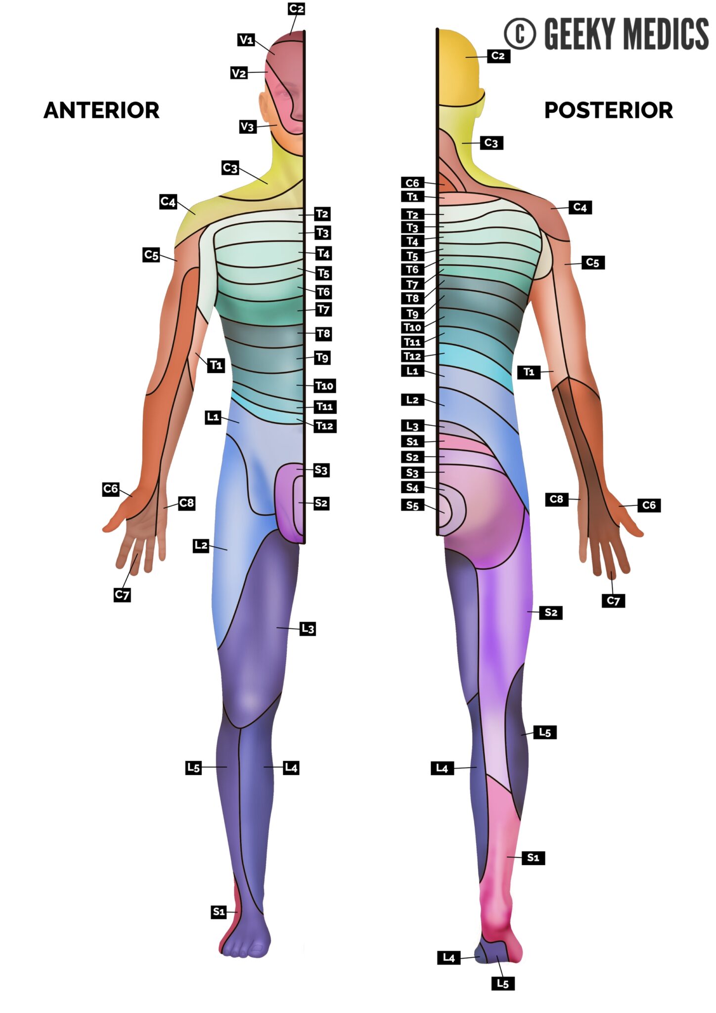
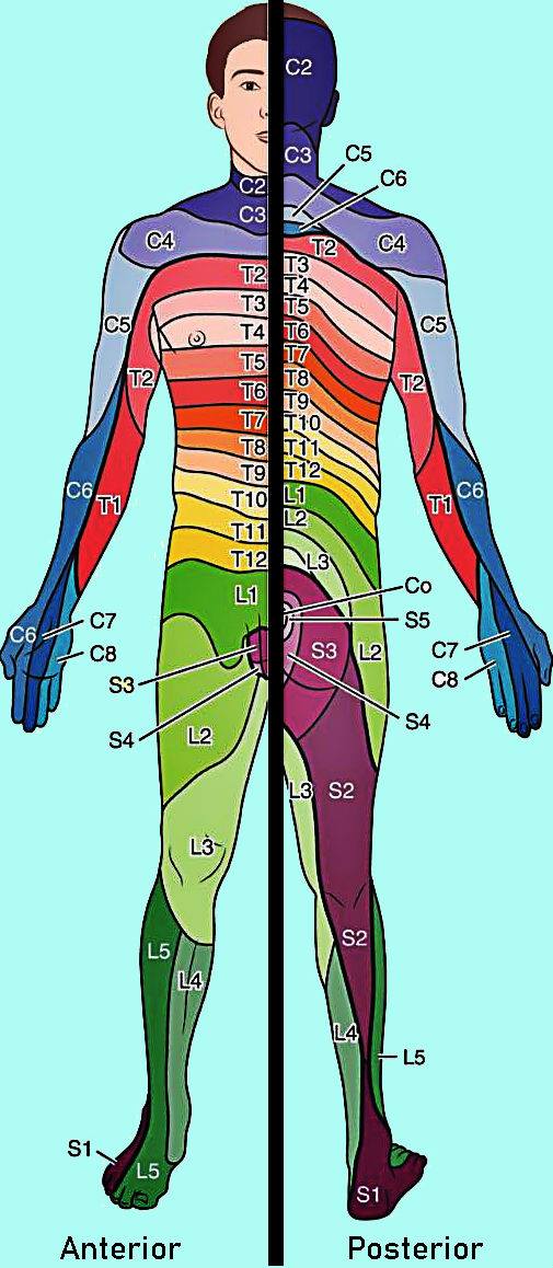
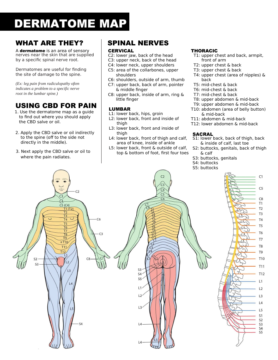
Dermatome Map:
-
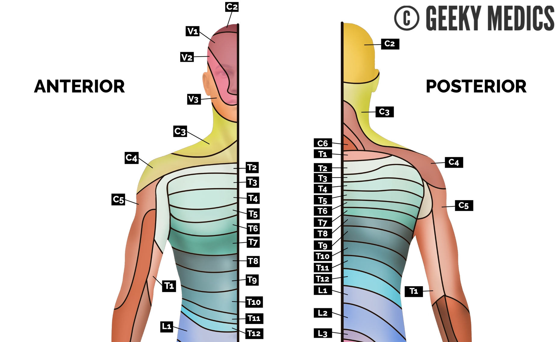
Cervical Dermatomes (C1-C8): These dermatomes cover the head, neck, shoulders, and upper extremities.
- C1: The back of the head.
- C2: The back of the head and the top of the neck.
- C3: The sides of the neck and the upper chest.
- C4: The upper chest and shoulders.
- C5: The lateral arm and the thumb.
- C6: The lateral forearm and the index and middle fingers.
- C7: The middle finger and the back of the hand.
- C8: The little finger and the medial forearm.
-
Thoracic Dermatomes (T1-T12): These dermatomes cover the chest, abdomen, and back.
- T1: The medial arm and the medial forearm.
- T2: The upper back and the axilla.
- T3-T6: The chest and the upper abdomen.
- T7-T12: The lower abdomen and the back.
-
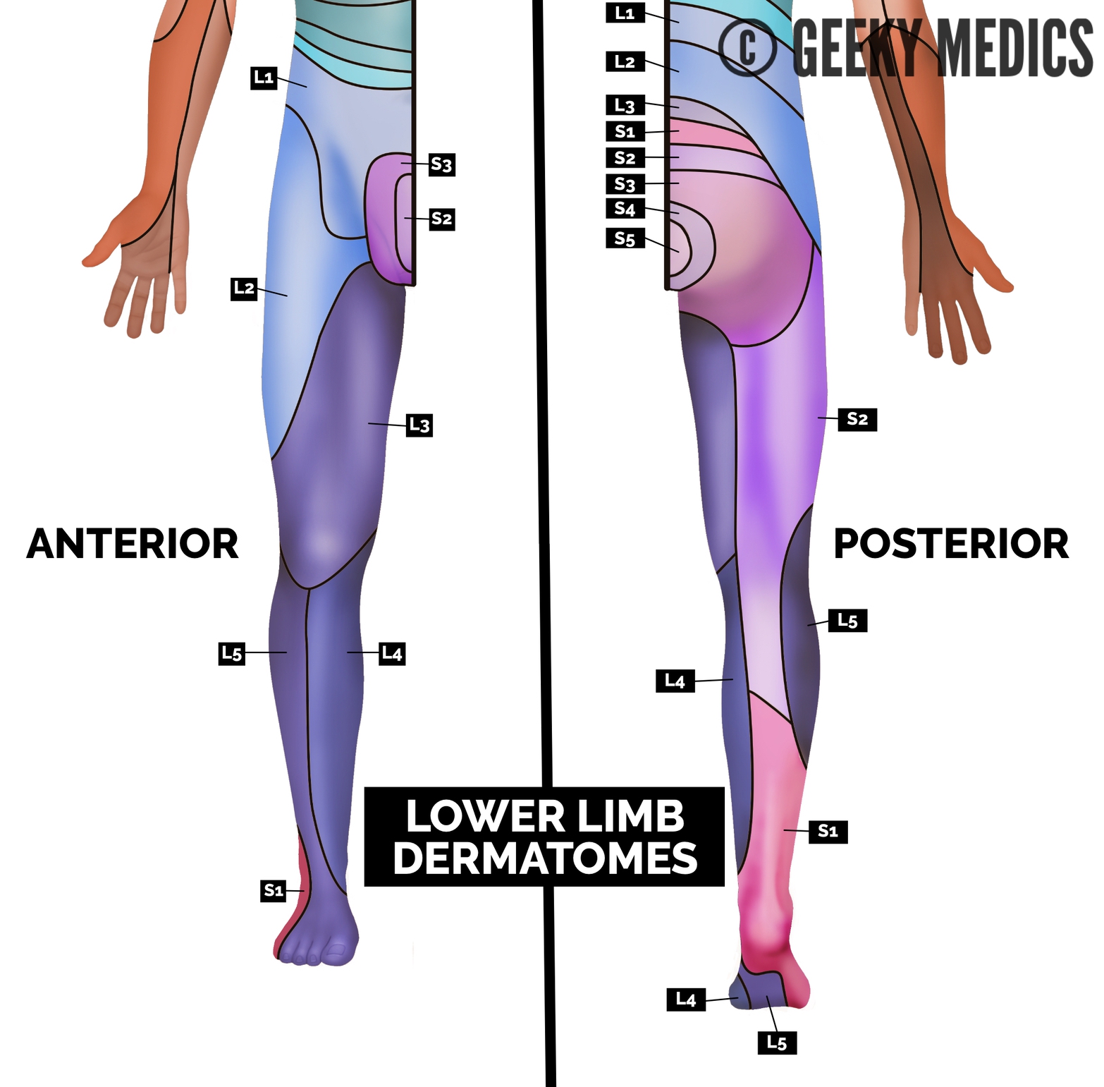
Lumbar Dermatomes (L1-L5): These dermatomes cover the lower abdomen, the groin, the front of the thigh, and the lower leg.
- L1: The lower abdomen and the groin.
- L2: The front of the thigh.
- L3: The front of the knee.
- L4: The medial calf and the medial ankle.
- L5: The lateral calf, the dorsum of the foot, and the big toe.
-
Sacral Dermatomes (S1-S5): These dermatomes cover the buttocks, the back of the thigh, the calf, and the foot.
- S1: The lateral foot, the heel, and the back of the thigh.
- S2-S4: The buttocks, the back of the thigh, and the posterior leg.
- S5: The perianal region.
Myotome Map:
-
Cervical Myotomes (C1-C8): These myotomes control the muscles of the head, neck, shoulders, and upper extremities.
- C1: Head flexion and extension.
- C2: Neck flexion and extension.
- C3: Shoulder elevation.
- C4: Shoulder abduction.
- C5: Shoulder abduction, elbow flexion, and supination.
- C6: Wrist extension and elbow flexion.
- C7: Wrist flexion and elbow extension.
- C8: Finger flexion and abduction.
-
Thoracic Myotomes (T1-T12): These myotomes control the muscles of the chest, back, and abdomen.
- T1: Intercostal muscles.
- T2-T12: Intercostal muscles, back muscles, and abdominal muscles.
-
Lumbar Myotomes (L1-L5): These myotomes control the muscles of the hip, thigh, and leg.
- L1: Hip flexion.
- L2: Hip flexion and knee extension.
- L3: Knee extension.
- L4: Ankle dorsiflexion and inversion.
- L5: Big toe extension and ankle dorsiflexion.
-
Sacral Myotomes (S1-S5): These myotomes control the muscles of the foot, ankle, and toes.
- S1: Ankle plantarflexion and eversion.
- S2-S4: Toes flexion and plantarflexion.
- S5: Perineal muscles.
Understanding the Maps: Key Considerations
-
Overlap: Dermatomes and myotomes exhibit some degree of overlap, meaning that multiple nerve roots may innervate the same area. This overlap provides redundancy and ensures that if one nerve root is damaged, other roots can compensate for its function.
-
Individual Variations: The exact distribution of dermatomes and myotomes can vary slightly from person to person. These variations are influenced by genetic factors and developmental processes.
-
Clinical Applications: The dermatome and myotome maps are essential tools in clinical practice. They help healthcare professionals understand the underlying neurological basis of various symptoms, guide diagnostic procedures, and formulate effective treatment plans.
Frequently Asked Questions (FAQs) about Dermatomes and Myotomes
1. What are the clinical implications of a dermatome or myotome deficit?
A deficit in a dermatome or myotome can indicate a problem with the corresponding spinal nerve root. This problem could be caused by various factors, including nerve compression, inflammation, or damage. The clinical manifestations of a dermatome or myotome deficit can vary depending on the location and severity of the nerve involvement. For example, a C6 dermatome deficit might present as numbness and tingling in the index and middle fingers, while a C6 myotome deficit might result in weakness in wrist extension.
2. Can dermatomes and myotomes be used to diagnose specific diseases?
While dermatomes and myotomes are not definitive diagnostic tools for specific diseases, they can provide valuable information that helps clinicians narrow down the possibilities. For example, a patient presenting with radiculopathy (nerve root compression) might exhibit characteristic sensory and motor deficits consistent with a specific dermatome or myotome. This information, combined with other clinical findings, helps in determining the underlying cause of the symptoms.
3. How can I learn more about my own dermatomes and myotomes?
There are various resources available to learn more about dermatomes and myotomes. You can consult with a healthcare professional, explore reputable medical websites, or refer to anatomy textbooks. Understanding your own dermatomes and myotomes can empower you to better understand your body and its neurological system.
Tips for Understanding Dermatomes and Myotomes
-
Visualization: Use anatomical charts and diagrams to visualize the dermatome and myotome maps. This visual representation can enhance your understanding of the nerve root distribution and their corresponding areas of the body.
-
Hands-on Exploration: Gently touch different areas of your skin and identify the corresponding dermatomes. This tactile exploration can help you grasp the concept of sensory innervation.
-
Muscle Movement: Experiment with different muscle movements and try to identify the myotomes involved in each action. This can improve your understanding of motor pathways and nerve root control.
-
Clinical Application: Observe healthcare professionals using dermatome and myotome maps in clinical settings. This exposure can provide valuable insights into the practical applications of these concepts.
Conclusion: A Deeper Appreciation for the Body’s Network
The dermatome and myotome maps serve as invaluable tools for understanding the intricate network of nerves that control our senses and movements. They provide a framework for diagnosing neurological conditions, evaluating muscle strength and sensory function, and guiding treatment strategies. By appreciating the complex interplay between the nervous system and the musculoskeletal system, we gain a deeper understanding of our own bodies and the remarkable mechanisms that govern our daily functions.


![Dermatomes and Myotomes [The Comprehensive Guide 2023]](https://physiohealthexpert.com/wp-content/uploads/2023/01/Dermatomes-and-Myotomes.jpg)

Closure
Thus, we hope this article has provided valuable insights into Unraveling the Body’s Network: A Comprehensive Guide to Dermatomes and Myotomes. We appreciate your attention to our article. See you in our next article!